Research laboratories
Applied Geophysics
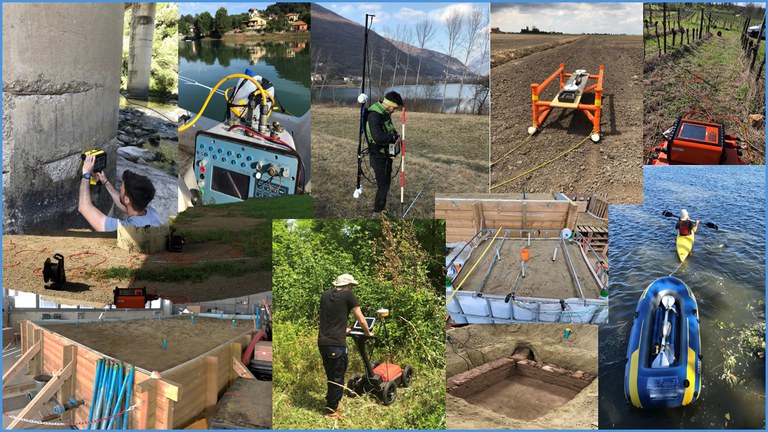
The Applied Geophysics Laboratory is equipped with a range of recently upgraded instruments. The laboratory's activities are focused on developing new solutions for geophysical characterisation and monitoring in order to actively contribute to various areas of applied geophysics research:
UrbanGeophysics
Hydrogeophysics
ArchaeoGeophysics
AgroGeophysics.
The Laboratory is equipped with the following devices:
- Geoelectricity and Induced Polarisation: Terrameter LT ABEM (48 electrodes with 10 channels)
- Ground-penetrating radar: IDS 400 MHz, C-True (IDS) 2 GHz, GSSI UtilityScan® DF (300–800 MHz) with GPS
- Geomagnetic: Overhauser GSM-19 from GEM System
- Passive Seismic survey: HVSR with Vibralog instrument with 3D seismic sensor with 2Hz frequency
- Active Seismic survey: 24-channel acquisition system (MASW and Refraction)
- Ultrasound: Pundit 200 from Proceq company
- Active and passive electrical methods for sample acquisition
- Multi-channel continuous electrical signal acquisition systems and current generators
The laboratory uses a research facility belonging to the Department to carry out experiments with quasi-real-life models. The facility consists of a 55 m³ tank filled with geological material and equipped with a permeameter system to simulate the aquifer. In addition, through a network of collaborations with research institutes (CNR) and national and international universities, the laboratory has access to other geophysical instrumentation facilities
Building F, Room F007
Referente: Prof. Enzo Rizzo
Atomic Physics

The atomic physics laboratory is a scientific facility of approximately 30 m² dedicated to studying physical phenomena within the field of atomic physics. In particular, it is equipped with optical and vacuum instrumentation that allows for experiments with magneto-optical traps on gases and the investigation of fundamental atomic transitions in alkali atoms at very low temperatures. The laboratory features large optical tables on which lasers and beam optics are installed, as well as control and management instrumentation such as spectrum analyzers and lock-in amplifiers. Preparatory measurements for experiments on unstable francium atomic beams are also carried out in this laboratory.
Buildings C, Room C219
Contact person: Prof. Luca Tomassetti
Birefringence Measurements
The birefringence measurement laboratory is located within a cleanroom of ISO Class 4, measuring 6.4 x 8.9 m², inside Building G. Its purpose is to measure the ellipticity induced by very small birefringences in both transparent samples and reflective surfaces. The cleanroom is maintained at a temperature of 23 ± 0.1°C and a controlled relative humidity of 56%.
An adjacent area measuring 2.5 x 9.0 m² houses the electronics operating the optical polarimeter. Inside the cleanroom, a granite optical bench measuring 4.8 x 1.5 x 0.5 m³ is supported by an isolation system with six degrees of freedom control. Ellipticity measurements can be performed at two wavelengths: 1064 nm and 1550 nm. The setup includes a structure supporting two rotating dipole permanent magnets, each rotating at approximately 10 Hz, producing a magnetic field B = 2.5 T with a length of 84 cm. These magnets are mechanically decoupled from the optical bench. A 2 cm diameter aperture allows light to pass through the magnetic field. When needed, the entire optical path can be placed under vacuum down to a residual pressure of approximately 10⁻⁷ mbar or operated in a controlled gas environment.
The polarimeter is also equipped with a Fabry-Perot interferometer to enhance sensitivity under certain measurement conditions, for example, magneto-optical effects in gases. The instrumentation includes two lasers, lock-in amplifiers, and spectrum analyzers.
Building G, Room G015
Contact person: Prof. Guido Zavattini
Coastal geomorphology COSTUF

The Ferrara Coastal Study Unit Laboratory (COSTUF) focuses mainly on coastal morphodynamics, including various types of environments such as urbanised and natural beaches, coastal dunes, mixed-grain beaches and wetlands in deltaic environments. Research has often been conducted as part of FP7 and H2020 projects supported by the European Community. COSTUF has also collaborated with private companies and public bodies such as ENI (the Italian oil company), Consorzio Venezia Nuova (local delegate for the Venice Water Authority), Thetis S.p.A. (environmental engineering company based in Venice), the Emilia-Romagna Region (regional authority based in Bologna, responsible for the Emilia-Romagna regional coastline), the Marche Region (regional authority based in Ancona, responsible for the Marche regional coastline), etc. The COSTUF group has often attracted visiting scientists from the United States, Australia and various European countries (France, Spain). The research group maintains a range of oceanographic equipment capable of collecting measurements of physical parameters in coastal and river environments. Thanks to ministerial funding from the Departments of Excellence, a Leica P30 Laser Scanner and a CK marine drone manufactured by Codevintec have recently been acquired. The laboratory is equipped with the following devices:
- Valeport Electromagnetic Current Meter;
- DLN 70 pressure transducer;
- Trimaran (Trimaran Ocean Science);
- RUNTI: Remote Unit for Nearshore Transport Investigation;
- RCM 9 Mk II acoustic current meter (AANDERAA);
- Turbidimeter OBS-3°;
- ADCP - Acoustic Doppler Current Profiler RDI Teledyne;
- Ohmex SonarMite echo sounder;
- Receivers Trimble GPS-RTK (R6 e R8);
- Current meter and wave meter (S4ADW InterOcean System);
- UAV - drone (quadcopter) DJI Phantom 3 Professional;
- Fluorescent tracers for sand;
- RFID radio trackers for pebbles and gravel;
- Video cameras (video surveillance);
- Codevintec CK-14 Marine Drone;
- Laser scanner Leica P30;
Building F, Room F008
Contact person: Prof.Paolo Ciavola
Detectors of elementary particles
Cryogenic Photon Detectors
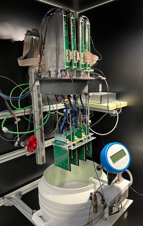 The cryogenic photon detector laboratory is an experimental area of around 50 m² located in Building G, with the capability for suspended load handling. The facility is equipped with instrumentation for the study and characterization of single-photon detectors across a range of temperatures from room temperature down to –200°C. It features both an automated system—allowing simultaneous study of 120 Silicon Photomultiplier (SiPM) sensors in parallel (named CACTUS)—and dedicated test benches for independent measurement of the fundamental parameters of these sensors.
Instrumentation includes low-noise source-meter units, digitizers, optical analyzers, and ultra-fast pulsed lasers, enabling comprehensive characterization of SiPM sensors. The laboratory also contains two large dark rooms (4 m³ and 2 m³, respectively) designed to house equipment that must be shielded from light during measurements.
Building G, Room G013
Contact person: Prof. Roberto Calabrese
DAQ Detectors
The DAQ Laboratory is primarily dedicated to the design, development, and testing of control, measurement, and data acquisition systems for experimental setups used in research and development activities for particle detectors. The laboratory’s equipment is based on oscilloscopes, CAEN and PICO digitizers, standalone National Instruments controllers, microcontrollers, and single-board computers such as Raspberry PI. Additionally, it features technical gas and water supply lines, making it a multidisciplinary facility where systems can be developed for general applications in physics, as well as in geology, architectural technology, and medicine.
Examples of developed systems include test setups for electronic measurement systems, standalone systems for test beams on detectors, long-term monitoring systems of environmental parameters for architecture applications, measurement systems for geophysics, and experimental support systems for high-energy physics experiments such as LHCb, DUNE, BESIII, etc.
The DAQ laboratory is also equipped with a laser cutter, sets of educational robotics/home automation kits, and a space arrangement suitable for educational activities aimed at university students, schools, and teacher training.
Building C, Roon C218
Contact person: Dr. Mirco Andreotti
Innovative Detectors Development
The “Innovative Detector Development” laboratory is located on the third floor of Building C. The laboratory is equipped with all necessary instrumentation for the characterization and deployment of various types of detectors. It features gas lines (with related alarms) connected to gas cylinders on the ground floor for innovative gas detectors; a dark room for testing photodetectors designed for medical physics and high-energy physics applications. Additionally, the lab includes scintillator bars connected to photomultiplier tubes (PMTs) or SiPMs, and commercial NIM electronics for trigger logic circuits, as well as oscilloscopes, computers, and power supplies for powering and controlling the systems.
Building C, Room C334
Contact person: Dr. Giuanluigi Cibinetto - Prof. Isabella Garzia - Dr. Giulio Mezzadri
Construction of Ultralight Detectors
The “Construction of Ultralight Detectors” laboratory is located on the ground floor of Building G, with the capability for suspended load handling. The laboratory houses all the equipment necessary for the fabrication of mechanical structures for ultralight gas detectors. Additionally, the lab is equipped with a compressed air line, granite tables, and various vacuum pumps for precision bonding and assembly operations.
Building G, Room G010
Contact persons: Dr. Giuanluigi Cibinetto - Prof. Isabella Garzia - Dr. Giulio Mezzadri
Photon Detectors Testing
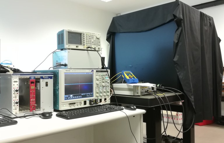 The photon detector testing and development laboratory is an experimental area of approximately 25 m² dedicated primarily to studying the fundamental characteristics of state-of-the-art photon detectors. The laboratory is equipped with two main scientific stations, each consisting of a stabilized optical bench, a large dark room able to house diverse instrumentation, an ultrafast pulsed laser with fiber optic output, and various control and management equipment suited for devices of different natures. The laboratory also hosts a remotely controllable grating monochromator with an output wavelength range spanning 300–1000 nm. It is equipped with high-efficiency light-blocking curtains, a dry air line, and a cooling chiller capable of maintaining mechanical parts at temperatures down to –10°C. The laboratory is mainly dedicated to quality testing of innovative photon detectors for high-energy physics applications.
Building C, Room C215
Contact person: Prof. Massimiliano Fiorini
ASIC
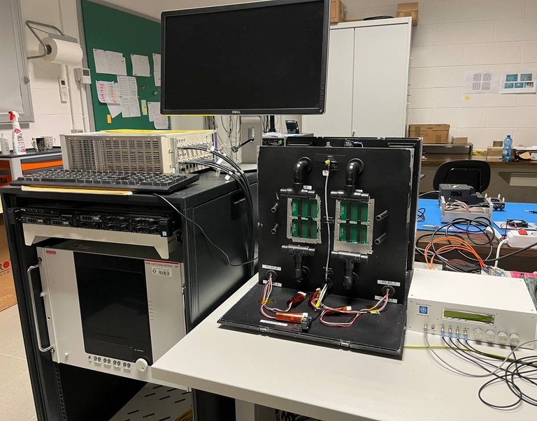 The ASIC laboratory is an experimental laboratory mainly dedicated to the characterization and study of ASICs designed and fabricated specifically for high-energy physics applications. This laboratory is equipped with cutting-edge technology that allows quality assurance testing of PCBs and ASICs. The laboratory has six independent workstations equipped with antistatic instrumentation (ESD), each outfitted with specific testing equipment such as source meter units, pick&place manipulators for micro-positioning integrated circuits, and control and management electronics. In particular, two workstations are equipped with a test system for CLARO chips, while another two are used for testing elementary cells housing the electronics and photodetectors of the RICH detector of the LHCb experiment. Additionally, one workstation is dedicated to the extensive characterization of the properties of the TIMEPIX4 ASIC.
Building G, Room C110
Contact person: Prof. Massimiliano Fiorini
Education Laboratories
Education Physics
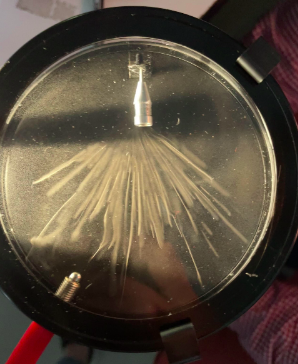 The Education Physics Laboratory is dedicated to teaching the scientific method and problem-solving from multiple perspectives, aiming to prepare future teachers to be critically proactive in teaching physics. The laboratory has three autonomous workstations, each equipped with different instruments, either in the form of "educational measurement kits" or custom-made setups. These allow students first to build and then independently and thoroughly experiment with various physical principles across mechanics, dynamics, fluid dynamics, thermodynamics, optics, magnetism, and modern physics in an interconnected and transversal way.
Experimental lessons for the "Physics Education" course are held in this laboratory. Additionally, postgraduate training courses are provided for secondary school teaching staff.
Buildings C, Room C317
Contact person: Prof. Giuseppe Ciullo
General Electronics and Measurement Systems
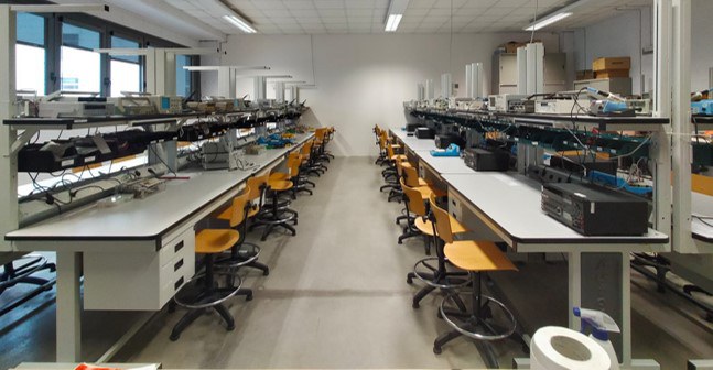 The General Electronics and Measurement Systems Laboratory is an educational laboratory dedicated to the study of analog and digital general electronics as well as acquisition and measurement systems. The laboratory has 20 independent workbench stations, each equipped with a construction kit for electronic circuits including passive and active components, a soldering station, measurement oscilloscope, and programmable microcontrollers.
The laboratory allows students to perform general electronics experiments starting from building simple discrete-element circuits on breadboards to designing and implementing more complex acquisition and measurement systems based on sensors and both standard and custom-built devices. The development of acquisition and measurement systems primarily relies on the use of Arduino microcontrollers, but projects also include the creation of onboard systems for smartphones.
The laboratory is mainly used for the “Electronics Laboratory” course for second-year students of Bachelro Degree in Physics, both during the semesters dedicated to digital and analog electronics and during the semester focusing on microcontroller programming and signal acquisition techniques.
Building F, first floor
Contact person: Dr. Mirco Andreotti
Modern Physics
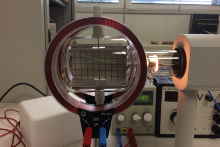 The Modern Physics Laboratory is an educational facility dedicated to the study of physical phenomena discovered since the 20th century, marking a significant break with the previous classical conceptual framework. The laboratory is equipped with various adaptable workstations where students can conduct numerous educational experiments on topics in atomic and nuclear physics.
Some of the key experiments possible include fundamental discoveries of physics over the past 100 years, such as: the Stefan-Boltzmann law for measuring the total radiation emitted by a black body, measurement of the speed of light using the superheterodyne method, the Franck-Hertz experiment on quantization, measurement of the photoelectric effect, Millikan’s oil drop experiment for measuring electron charge, atomic spectral measurements, studies on the behavior of cathode ray, study of the Hall effect, the Zeeman effect and measurements of NMR and ESR, X-ray measurements and Moseley’s law, the Compton effect, Rutherford’s experiment, measurement of particles and tracks in Wilson’s cloud chamber, radioactivity measurements.
The laboratory is primarily used for the "Modern Physics Laboratory" course for undergraduate Physics students.
Building C, Room C013
Contact person: Prof. Giuseppe Ciullo
Radiation-Matter Interaction
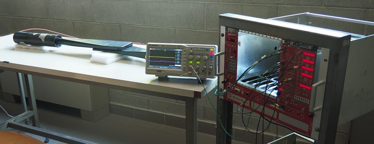 The Radiation-Matter Interaction Teaching Laboratory is dedicated to conducting experiments for the detection of elementary particles, allowing students to experience the fundamental phenomena governing interactions between radiation and matter. The laboratory is equipped with six workstations, each fitted with typical instrumentation from nuclear and subnuclear physics experiments, including scintillators and photomultiplier tubes, discriminators, signal shapers, ADC and TDC modules, logic gates, and digitizers.
Each workstation allows groups of 2–3 students to work independently on phenomena related to nuclear and subnuclear physics, particularly interactions between particles and matter.
The laboratory hosts the experimental component of the "Radiation-Matter Interaction Laboratory" course for the Physics degree program and the "High Energy Physics Laboratory" course for Master's students in Physics. Demonstrations are also carried out during outreach and public engagement events.
Building G, Room C116
Contact person: Prof. Roberto Calabrese
Mechanics and Dynamics
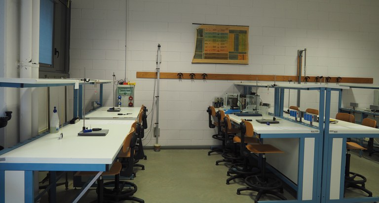 The Mechanics and Dynamics Laboratory is an educational facility organized into 20 independent workbench stations, each equipped with different instrumentation allowing experiments on mechanics and body dynamics topics. The laboratory supports experiments with simple pendulums, Kater’s pendulum, torsion pendulums, springs and dynamometers, guides and timing gates for free-fall experiments, inclined planes, thermometers, thermostatic baths, calorimeters, function generators, and oscilloscopes for measuring wave propagation speeds in air.
The laboratory can accommodate up to 40 people simultaneously, who can perform experiments individually or in small groups of 2-3. It is primarily used for the experimental part of the "Physics Laboratory with elements of statistics and computer science" course dedicated to first-year undergraduate Physics students.
Building F, Room F005
Contact person: Prof.ssa Eleonora Luppi
Optics
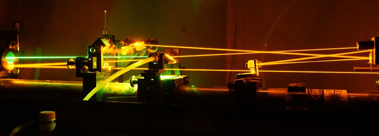 The Optics Teaching Laboratory is a modern facility designed for teaching the main laws of classical and quantum optics. The laboratory is equipped with 12 independent optical benches, each fitted with its own diode laser, a set of optical mounts, mirrors, lenses, polarizers, and light detectors. Each station can be used individually or by small groups of 2-3 students, allowing them to independently experiment with numerous optical phenomena including the laws of reflection and refraction, the thin lens formula, diffraction and interference of light, and linear and circular polarization phenomena.
The laboratory hosts the experimental component of the "Optics Laboratory" course for third-year Physics undergraduates, as well as other teaching activities and experiences related to optics.
Building G, Roon C113 and G114
Contact person: Prof. Roberto Calabrese
Gemmology
The Laboratory is equipped with the main instruments for gems identification, including stereomicroscopes, polariscopes and refractometers, which can be used for teaching, research, dissemination and third mission activities. The laboratory has a range of instruments for research purposes: digital stereo microscopes with transmitted and reflected light, conoscopes, refractometers, polariscopes, dicroscopes, UV lamps, contact liquids, hydrostatic scales, normalised light lamps, cubic zirconia masterstones, gemmological data comparison tables, main cut comparison tables, and main gem inclusion comparison tables.
In particular, the laboratory also has the following instruments:
- Zeiss microscope: SteREO Discovery V12 body with motorised optical zoom; lower base for transmitted LED light (bright field, oblique, dark field); LED illumination for reflected light with adjustable fibre arms; 1.25X objective lens (magnification from 10X to 120X with 10X eyepieces) • PL 10x eyepieces; 8 mpx full HD 4K colour microscopy camera • Transmitted light and reflected light polarisation.
- Motic SMZ-143-N2TG digital gemmological stereo microscope with CCD camera. Incident light and transmitted light 3W LED with adjustable intensity; zoom magnification from 10x to 40x; 45° inclined binocular head, 360° rotatable with dioptre adjustment.
Building B, Room PT
Referente: Prof.ssa Annalisa Martucci
Geochemistry
Cutting-grinding
The main activity, which takes place in two separate areas, is the production of various types of end products (granules, powders, concentrates of specific minerals, etc.), normally through a process of sample reduction by cutting, crushing and grinding. The activities commonly carried out are:
- Reduction to coarse/fine grain size (5 mm – 100 microns) using crushers and mortars, for sieving using vibrating screens and/or mineral separation (Frantz, hand-picking, inclined table, etc.).
- reduction to powder (50-5 microns) using mills in order to perform total rock analysis (XRF, XRD, ICP-MS, etc.)
The laboratory contains the following devices :
- Reinforced steel jaw crusher: suitable for rapid crushing of materials ranging from medium to extreme hardness, as well as friable and plastic-hard materials. Some examples: various types of rock (granite, basalt, limestone, etc.), concrete, cement clinker, gravel, slag, metal ores, glass, etc. The achievable output fineness is normally <5 mm. The maximum feed opening is 90 mm.
- Disc mill (Retsch RS100): allows ultra-fine grinding (<10 microns) of medium-hard or hard brittle materials, in dry or wet condition. The powders obtained in this way can be used for chemical and/or mineralogical analysis (XRD). The previously crushed material is ground in an agate grinding apparatus consisting of a jar and several rings, in order to ensure negligible contamination of the sample for subsequent chemical analysis. The 100 ml volumetric capacity allows sufficient material to be obtained in a single pass for subsequent analysis, as well as excellent homogenisation of the sample.
- Larmann pestle mill: allows grinding to various degrees of fineness (50-2 microns) of any type of material (both fragile and plastic) of a mineral or organic nature. Grinding can be done either dry or wet. The powders obtained can be used for chemical and/or mineralogical analysis (XRD). The grinding of the previously crushed material takes place in a grinding apparatus made entirely of agate. The apparatus consists of a jar and a pestle driven by an electric motor. Unlike other devices, this instrument allows the grinding of very small quantities of material and is therefore ideal for use in cases where only very small quantities of sample are available.
Building B, Room PT
Contact persons: Prof. Emilio Saccani, Dr. Renzo Tassinari
Mineral separation
his laboratory allows for the mineralogical separation of rock samples, which is useful for subsequent crystallochemical, geochemical or petrological analyses. Minerals with a grain size of less than 200 μm and different magnetic susceptibilities can be separated using the Frantz magnetic separator (S.G. Frantz Co. L-1). This is an electromagnetic device that allows the separation of different mineralogical phases according to their magnetic susceptibility and density. For optimal operation, the material introduced into the device must have a well-defined grain size, which can be obtained by sieving. The device, through continuous adjustment of various parameters (grain size, longitudinal and lateral inclinations of the slide, magnetic force), and several passes under different conditions, can be used to achieve different degrees of mineral separation. From simple separation of magnetic/non-magnetic minerals, to the separation of groups of minerals, to that of individual mineral phases.
Building B, Room PT
Contact persons: Prof. Emilio Saccani - Prof.ssa Costanza Bonadiman , Dr. Renzo Tassinari
Geochemical Preparation
The laboratory is dedicated to the preparation of rock, soil, water and sediment samples for geochemical analysis, as well as the quantitative determination of certain elements (e.g. volatile elements, CO2, organic matter). The activities carried out are: acid attack of lithoid material powders, determination of loss on ignition at 1000°C, determination of CO2 content in rocks (calcimetry), determination of organic matter by calcination at 500°C, production of glass “beads” from lithoid samples for XRF analysis, removal of organic matter and soluble salts from sediment samples by washing with hydrogen peroxide at 130 vol. Determination of loss on ignition (calcination). The samples are calcined to determine their loss on ignition (L.O.I.) by placing them in an oven at 105 °C for about 4 hours, and then in a muffle furnace at 1050 °C for about 5 hours.The procedure is used to determine the content of volatile elements in minerals. Determination of the amount of organic matter in soils and sediments. The samples are calcined at 500°C in a muffle furnace for about 5 hours because, at that temperature, the organic matter is volatilised. Determination of the CO2 content (calcimetry) in various materials using a simple volumetric calcimeter. The samples are reacted with HCl, which produces CO2. Preparation of glass discs (“beads”) for the analysis of major elements in XRF. The activity consists of melting sample powder using lithium tetraborate (Li2B4O7) as a flux with a dilution of 1:10. Melting is carried out using an Equilab FX1 induction melting furnace (maximum temperature reached approximately 1000°C).
The laboratory contains the following devices :
- Oven with variable temperature from 50°C to 350°C (to be verified), programmable working times and temperature rise gradient.
- Muffle furnace for high temperatures (350°C-1050°C) programmable for maximum temperature, working times and temperature rise gradient.
- Equilab FX1 pearling machine equipped with melting equipment (crucibles + plates) in both platinum and zirconium. The instrument can melt rock powders at temperatures up to 1000°C using an induction oven. All stages of the procedure (heating, stirring of the melt, casting and cooling) are automatic, programmable and controlled by a dedicated computer. The instrument is also capable of performing alkaline fusions for ICP-MS analysis.
- Simple volumetric calcimeter consisting of a sample-acid reaction ampoule, a graduated tube for measuring the volume of CO2 produced by the reaction, and an expansion vessel.
- Two heating plates with various sample holder accessories used for acid attacks.
- Water purifier that produces 18 MΩ water for geochemical analysis. Fume hoods, one of which is designed for use with hydrofluoric acid.
- Special cabinets for storing acids and flammable materials.
- Special benches for chemical laboratories.
Building B, Room PT
Contact person: Prof. Emilio Saccani - Prof.ssa Costanza Bonadiman -Dr. Renzo Tassinari - Dr. Massimo Verde
Inductively Coupled Plasma Mass Spectrometry (ICP-MS) and Ion Chromatography
Inductively coupled plasma mass spectrometry (ICP-MS) is recognised as a key technique for multi-element analysis at ultra-trace concentration levels in a wide variety of applications. This is due to its high speed, analytical sensitivity and ability to achieve detection limits generally below ng/L for most elements in the periodic table. With triple quadrupole technology, such as the iCAPTQ ICP-MS model used in this laboratory, the constant reduction of interfering species is optimised regardless of the sample matrix, thus allowing for a wider range of investigations. The reduction of spectroscopic interference is achieved through the use of a multipolar collision/reaction cell (CRC) and a wide variety of gases. The laboratory's activity consists of determining trace and ultra-trace elements in various types of samples suitably prepared in solutions obtained from acid attack. Several techniques are used:
- Quantitative analysis of solutions using external calibration curve
- Quantitative analysis of solutions using internal calibration curve by standard addition method
- Use of internal standards to compensate for variability during measurement and variations in the sample matrix
The instrument used is an ICP-MS-QQQ spectrometer, model iCAP-TQ, equipped with: chiller, pump, autosampler, software, PC, monitor and printer.
Ion chromatography spectrophotometry is based on the chromatographic separation of cations and anions using cation or anion exchange columns. The individual analytes are eluted in succession and determined by a conductometric detector after chemical or electrochemical suppression of the electrical conductivity of the eluent. The concentrations are obtained by integrating the areas of the individual chromatographic peaks and comparing them with calibration curves obtained by injecting solutions of known concentrations within the analytical investigation range. Qualitative and quantitative geochemical analyses of geological samples (rocks, minerals and natural waters).
The instrument used is an ICS-1000 DIONEX-THERMO SCIENTIFIC ion chromatograph equipped with an AS40 autosampler.
Building B, Room PT
Contact persons: Prof. Emilio Saccani - Prof.ssa Costanza Bonadiman- Dr. Renzo Tassinari
Stable Isotope Spectrometry
Isotope ratio mass spectrometry (IRMS) is an advanced and specialised technique of mass spectrometry designed to accurately measure the relative abundances of stable isotopes of various elements such as carbon (C), hydrogen (H), nitrogen (N), oxygen (O) and sulphur (S). This method provides a detailed and in-depth understanding of various natural and anthropogenic processes and phenomena, finding application in many fields of research and industry. Thanks to its ability to provide detailed and accurate information on isotopic abundances, it allows complex issues in the environmental, palaeoenvironmental, agricultural, forensic, archaeological and many other fields to be resolved, contributing significantly to our understanding of the natural world and its evolution over time, as well as human activities.
The laboratory contains the following devices:
- CN, EA elemental analyser (Elementar) IRMS mass spectrometer (Isoprime 100)
- CNSHO, EA PYRO-Elementar elemental analyser and IRMS precisION-Elementar mass spectrometer
- IRMS mass spectrometer coupled with specific isoflow peripheral device for isotopic analysis of carbonates
- Laser spectrophotometer for isotopic analysis of oxygen and hydrogen
- Los Gatos Research (LGS) LWIA 24 used is based on the CRDS (Cavity Ring Down Spectroscopy) technique, a laser spectroscopy technique based on Lambert-Beer's law, which relates the extinction time of a laser beam to the concentration of isotopic species in a vaporised water sample. This is essentially absorption spectroscopy, in which a source emits radiation (IR, UV-visible), and an analyte (atom or molecule) absorbs at a particular frequency determined by its energy configuration (atom) or by the type of bonds/masses present (molecule). It is therefore possible to obtain absorption spectra to obtain a qualitative description of the sample and, from the intensity of the absorption, to trace the concentration of the analyte using Beer's law. The most recent development, cavity ring-down spectroscopy, has made it possible to achieve a level of accuracy that allows it to be used for analysing the isotopic composition (18O/16O, 2H/1H) of water.
Building B, Room PT
Contact persons: Prof. Gianluca Bianchini - Prof. Gianluca Frijia
Geological Mapping - CARG
This laboratory is dedicated to geological mapping, consultation and preparation of geological maps at various scales and, in particular, to the creation and coputerisation of geological sheets at a scale of 1:50,000 relating to the CARG (CARtografia Geologica) project, in which the research group has been involved for many years. Specifically, the laboratory is equipped with workstations for digital cartography in a GIS environment and a rich collection of topographic, geological and geothematic maps.
Building B - Third Floor
Contact persons: Prof. Piero Gianolla - Prof. Michele Morsilli - Prof. Gianluca Frija
Geothermal energy
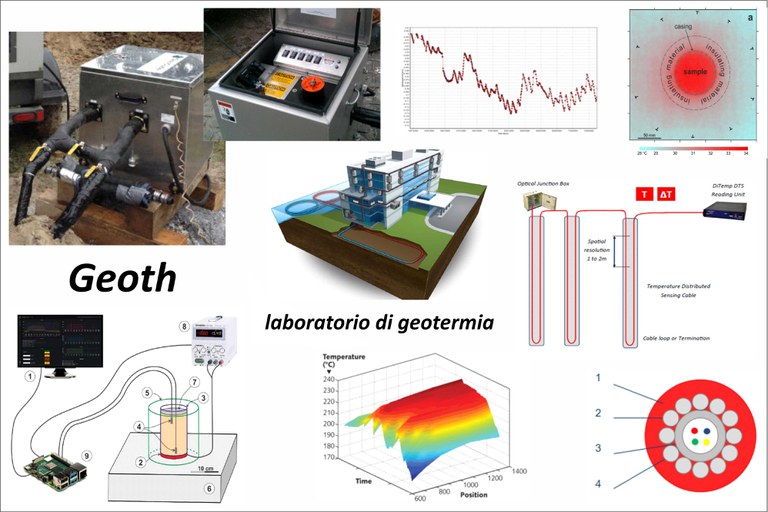
The geothermal laboratory allows research and development activities to be carried out in the field of renewable energy, with a particular focus on heat transfer in the subsoil (soil and aquifers) in different geomaterials and on varying the initial conditions (e.g. degree of saturation or presence of moving groundwater) and boundary conditions (e.g. external temperature).
The laboratory consists of various experimental devices that measure the thermal conductivity of soils and heat accumulation on a) samples of loose materials (lab-GRT), b) in situ inside wells of various types (field-GRT) and c) on rocks both in the field and in the laboratory (fast-GRT). The laboratory also has equipment for continuous temperature measurements using fibre optics with measurements every 2 m (Geothermal Monitoring). The laboratory also has instruments for measuring temperature in wells to a depth of 200 m, as well as a groundwater level meter and a multi-parameter sensor (pressure transducers, T, EC) for measurements up to 100 m. Finally, it is also possible to perform static and dynamic thermal analyses of surfaces using a thermal imaging camera (ThermoCam-Monitoring).
Building B and Building F
Contact persons: Prof. Riccardo Caputo - Prof.ssa Dimitra Rapti
Interferometry
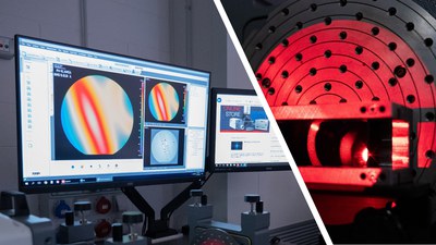
The Interferometry Laboratory enables quantitative and non-destructive morphological measurements on a wide range of samples of various sizes, shapes, and roughness levels. By exploiting the interference phenomenon of visible light, the profile along the observation axis is reconstructed with nanometric resolution. These maps allow the control of shape errors for lenses and mirrors, the definition of surface roughness, and the measurement of thicknesses of transparent thin films.
The instrumentation includes two complementary interferometers: the Zygo VeriFire HDX, with a large field of view (⌀ 200 mm) for large polished surfaces, and the Zygo NexView NX2, which achieves magnifications up to 100x and measures non-polished surfaces.
The activity mainly focuses on characterizing the curvature of curved crystals used as deflectors of high-energy particle beams in accelerators such as LHC at CERN, exploiting the channeling phenomenon. Additionally, the precision of feedback achievable with interferometric techniques has been utilized in component assembly, allowing alignment control between elements with precision better than 100 microradiants.
Building G, Room G006
Contact persons: Dr. Andrea Mazzolari - Dr. Marco Romagnoni
Magnetism
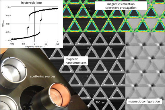 This research area focuses on the magnetic properties of nanostructured systems, in the form of thin films (thickness ~10 nm), both continuous and organized in periodic structures, and magnetic nanoparticles (diameter ~10 nm). Activities in the group include both experimental and theoretical work. From the experimental point of view, the Department is equipped to prepare samples containing nanostructures using a thin film growth system. Magnetic property analyses of the samples can be performed using a MOKE magnetometer and a SQUID magnetometer. The morphology and magnetic configuration of the samples can also be studied through magnetic force microscopy. Furthermore, compositional and structural characteristics of materials, as well as magnetic and non-magnetic phase transitions, can be investigated using scanning calorimetry and thermogravimetry.
The theoretical studies focus on analyzing the propagation of spin waves within nanostructures, employing micromagnetic simulation methods. These simulations utilize computing resources provided by the group and Department, including the high-performance computer cluster COKA as well as the Leonardo supercomputer at CINECA.
Building C, Rooms C004 - C014 - C015 - C016 - C020
Contact Persons: Prof. Diego Bisero - Prof. Lucia Del Bianco - Prof. Loris Giovannini - Prof. Federico Montoncello - Prof. Federico Spizzo
Micropalaeontology
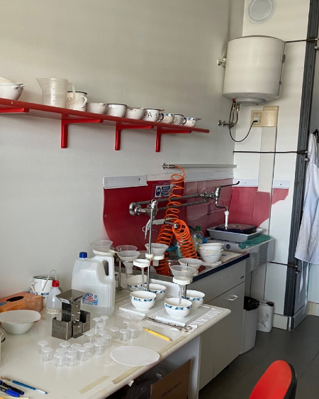
In this laboratory, rock samples are prepared in order to obtain washing residues from which microfossils can be isolated and identified for micropalaeontology and palaeoclimatology research. The procedures involve breaking down the samples using various types of agents (e.g. H2O2, Neosteramine) and washing them in running water with 63 and 38 micron sieves. This produces washing residues in which the microfossils are concentrated. Once dried in an oven, the washing residues are placed in special containers from which they can be placed on trays and identified under a stereomicroscope. The sieves are immersed in a methylene blue bath to stain any microfossils trapped in the mesh and prevent contamination. The laboratory is equipped with an oven for drying samples and washing residues, a balance, a press for fragmenting the most lithified rock samples, an ultrasonic tank for further cleaning of microfossils, a splitter for separating the residues into equal parts, and compressed air for cleaning the sieves. The laboratory is used by lecturers, PhD students, research fellows (for research), students (for degree theses) and 150 hours student assistants.
Building B, Room: B308
Contact person: Prof.ssa Valeria Luciani
Medical Physics
The teaching and research activities of the Medical Physics group, rwhich concern applications in diagnostic radiology, nuclear medicine, and biophysics of blood circulation, make use of various specialized laboratories within the Department.
Some of the experimental and teaching activities involving radiogenic sources also take place at the LARIXinfrastructure.
Eco-Fluidodynamics
The Eco-Fluidodynamics Laboratory is essential for the research activities of the Medical Physics group in the field of blood circulation biophysics. This laboratory is equipped with instruments for evaluating blood flow, including a portable ultrasound device for diagnostic imaging, several pumping systems for hemodynamic circuits that reproduce the circulatory system, and phantoms—both commercial and custom-developed—to simulate the anatomical region of interest. This equipment is primarily used to investigate the phenomenon of cerebral venous return, that is, to study the mechanisms ensuring blood flows back from the brain to the heart, as in the Drain Brain 2.0 experiment funded by the Italian Space Agency (ASI), which will be carried out on the International Space Station (ISS). The laboratory also hosts the device developed with the WISE, project: a wireless electronic system capable of synchronously detecting heart electrical activity and skin deformations due to blood pulsation. Additional work includes the development of wearable devices for monitoring the rehabilitation of cardiac patients, thanks to a research project funded by INFN (EPISE) conducted in collaboration with the Biomedical Studies Center applied to Sport at UNIFE.
Building C, Room C312
Contact persons: Prof. Angelo Taibi - Dr. Antonino Proto
Detectors for Medical Physics
The laboratory is equipped with the instrumentation required for the development and characterization of innovative detectors for ionizing radiation, particularly in the context of radioactivity measurements and imaging detectors for nuclear medicine and radiology. The laboratory has developed a beta radiation counter using the TDCR technique for the absolute measurement of radioactivity in liquid solutions. This device is employed to quantify the activity of radiopharmaceuticals produced by accelerators for use in nuclear medicine. There are also prototypes of detection systems for SPECT and PET, used to study the image quality performance of innovative detectors in preclinical research. In addition, various state-of-the-art workstations provide the computing resources necessary for tomographic image reconstruction and artificial intelligence applications.
Buildiing C, Room C313
Contact person: Prof. Giovanni Di Domenico
Computing for Medical Physics
The laboratory is used for activities concerning the application of computational methods in medical physics, such as the development and use of Monte Carlo particle tracking codes (on both CPU and GPU), the development of artificial intelligence algorithms, and the processing of large image datasets. The laboratory is equipped with two multiprocessor workstations featuring state-of-the-art graphics cards optimized for artificial intelligence. In addition, there are workstations available for remote access to two dedicated computing servers with 32 and 96 cores, respectively.
Building C, Room C314
Contact persons: Prof. Angelo Taibi - Dr. Gianfranco Paternò
Teaching Laboratory of Detectors in Medical Physics
At the Teaching Laboratory of Detectors in Medical Physics, activities include spectroscopy of X-ray and gamma radiation sources, as well as measurements for characterizing detectors used in diagnostic imaging. Many of the activities are conducted for educational purposes or as part of outreach and orientation initiatives aimed at high school students.
The laboratory is equipped with a complete spectroscopic chain based on sodium iodide (NaI) scintillation detectors coupled with photomultiplier tubes, signal readout electronics, and digitization systems. There is also a light-tight chamber equipped with servo-controlled movers used for characterization measurements and testing of imaging detectors (such as CCD cameras and photon-counting detectors), along with an optical bench with adjustable magnification for optical acquisitions.
Building C, Room C316
Contact persons: Prof. Giovanni Di Domenico - Dr. Paolo Cardarelli
Microscopy
This laboratory is dedicated to microscopic observation, both on isolated material and thin sections. It is used by lecturers, PhD students and research fellows for research purposes, and by students for analyses related to their bachelor's and master's theses. Specifically, the laboratory is equipped with five stereomicroscopes for the observation of isolated specimens and four transmitted light microscopes with different magnifications - up to 1000X - for the observation of thin sections.
The first group includes:
- Leica M 165 C. This device, the most powerful in the range, is equipped with a video camera and allows high resolution for thin section photos and isolated material.
- Zeiss STEMI 508 one of which has a video camera.
- Leica M80
The second group includes:
- Leica DM 2700 P
- Zeiss dual vision 47 30 34-9902. This instrument allows two operators to directly compare their observations.
- Zeiss 47 34-9902
- Olympus UTV1 INV. 000071B6
In addition, the laboratory is equipped with several drawers for storing samples.
Building B, Room: B311
Contact person: Prof.ssa Valeria Luciani
Nuclear Technologies Applied to the Environment
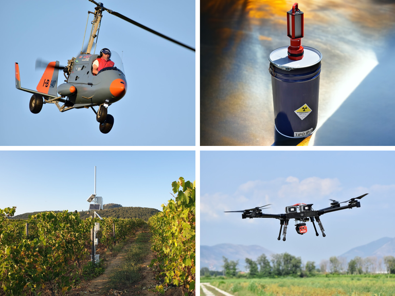 The Laboratory of Nuclear Technologies Applied to the Environment specializes in advanced technologies for detecting environmental radioactivity and applications in precision agriculture. It is equipped with scintillation and semiconductor detectors to detect and quantify natural and artificial radionuclides. In situ measurements use gamma spectrometers with CeBr3 and NaI(Tl) crystals, which can be mounted on drones and experimental aircraft like RadGyro, an autogyro developed for multiparametric surveys. Portable systems integrate these detectors for characterizing medical waste and materials contaminated with artificial radionuclides or Naturally-Occurring Radioactive Materials (NORM).
Laboratory measurements are performed with the MCA_RAD system, using shielded coaxial High Purity Germanium (HPGe) detectors that allow automated, high-resolution spectral acquisitions on solid or liquid samples.
In precision agriculture, the laboratory uses high-resolution RGB and multispectral cameras to monitor soil and vegetation cover. The data collected enable detailed analysis through algorithms that evaluate plant spectral signatures to identify health and diseases, supporting decision-making for agronomic interventions.
The laboratory also possesses a computing cluster for developing artificial intelligence algorithms to enhance data analysis, as well as 3D printers for creating customized detection systems tailored to specific project needs
Building C, Room C012 and C001
Contact person: Prof. Fabio Mantovani
Palaeobiology of calcareous algae and macroforaminifera
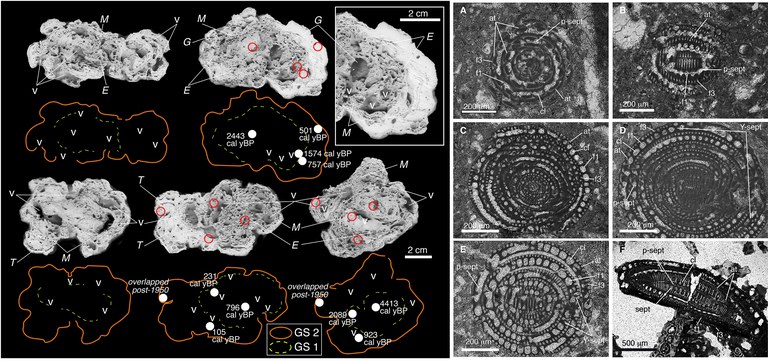
We study the evolution of marine ecosystems of carbonate and mixed siliciclastic-carbonate platforms over large time scales (Mesozoic-Holocene) by evaluating palaeoecological dynamics and palaeobiogeographical models.
Palaeoecological and palaeoenvironmental interpretations, dynamics and structures of palaeocommunities of calcareous algae and benthic macroforaminifera are obtained by combining palaeontological surveys, analytical methods and palaeobiological analyses (systematics, morphology, structure and architecture, taphonomy).
Laboratory research is carried out using incident light stereomicroscopy (Leica M50; zoom magnification 0.63x–4x) and transmitted light stereomicroscopy (Leitz Diaplan; magnification with 2.5x–100x lenses).
Building B, Room B206
Contact person: Prof. Davide Bassi
Palaeoclimatology and isotopic stratigraphy
The laboratory is equipped with state-of-the-art instruments for the preparation and petrographic and geochemical characterisation of carbonate matrices.
The laboratory includes:
- PrecisION IRMS mass spectrometer (Elementar) coupled with specific isoFLOW peripheral device (Elementar) for isotopic analysis of O and C in carbonate matrices. The instrument can be used for the reconstruction of environmental and diagenetic processes and for stratigraphic studies with implications, for example, in the reconstruction of palaeotemperatures and high-resolution stratigraphic correlations.
- Cathodoluminescence model CL MK 5: CL is used for diagenetic and petrographic studies. It can be used to highlight zoning and other microstructures in individual crystals and rocks that would otherwise be invisible, and is useful for characterising stone materials for industrial and archaeological purposes.
- MicroMill ESI autosampler: Computerised system consisting of a microscope combined with a micro-drill for high-precision sampling of solid materials. The instrument can be used to take micro-samples from stone materials (e.g. rocks, concrete, ceramics, etc.) for further analysis.
- WXTS3DU precision microbalance: Precision balance accurate to six decimal places for weighing powders intended for geochemical analysis. The instrument is used to accurately determine the initial weight of a sample subject to further analysis and the change in weight after any treatment.
- Georadis GT-32 Gamma Ray Spectrometer: Instrumentation for measuring natural gamma dose rates (Gy/h) and spectral measurements with determination of K, U, Th concentrations (%, ppm, ppm) for stratigraphic and environmental purposes.
Building B, Room PT
Contact persons: Prof. Gianluca Frija - Prof. Michele Morsilli
Thermal Analyses
The Thermal Analysis Laboratory is dedicated to the study of the chemical-physical transformations of powdered, mineralogical, synthetic inorganic and organic samples (building materials, ceramics, composites, etc.) subjected to controlled thermal cycles. It allows the determination of mass variation (wt% or in mg), characteristic transformation temperatures, kinetics and reaction mechanisms, exo-endothermic effects, glass transitions and crystallinity. The laboratory is equipped with interchangeable sample holders (TG, TG-DSC, TG-DTA, etc.) and it allows the simultaneous execution of thermogravimetric (TG) curves, differential thermogravimetric (DTG) curves, differential thermal analysis (DTA) and differential scanning calorimetry (DSC) curves simultaneously (STA/TG-DSC balance, STA409PC NETZSCH). Analyses can be conducted from room temperature up to 1500°C, in an air or nitrogen atmosphere. The instrument is coupled with a Micro-Gas Chromatograph (GC-MS) to detect the gases evolved and thus identify the individual components, correlating them precisely, at the time scale, to each signal of the thermoanalytical curve. The laboratory also has a controlled anaerobic atmosphere chamber (glove box) for applications requiring a confined space and/or protection of the environment or product. It can also be used in an inert gas atmosphere to protect the material under study. In detail, the Laboratory has the following equipment:
- Thermogravimetric Balance – Simultaneous Thermal Analysis/Differential Scanning Calorimetry (STA/TG-DSC), STA409PC Luxx, NETZSCH. Temperature range: RT-1,650°C. TG resolution: 2 µg; DSC resolution < 1 µW; Atmosphere: inert, oxidising, reducing, static, dynamic
- Agilent 490 Micro GC, with four-channel configuration, for analysing permanent gases, paraffins/olefins, carbon dioxide and hydrogen sulphide
- Glove Box Hood, Plas-Labs, Inc. TM Ambidextrous white Hypalon gloves for superior resistance to chemicals and UV rays. Transparent transfer chamber 12‘ long and 11’ diameter (lD). Optically transparent acrylic top. Adjustable vacuum gauge on transfer chamber. Four taps with grounding key for purging.
Building B, Room: PT
Contact persons: Prof. Giuseppe Cruciani - Prof.ssa Annalisa Martucci
Sedimentology
The sedimentology laboratory of the Department of Physics and Earth Sciences – University of Ferrara is equipped to perform textural and physical analyses of geomaterials from various natural or anthropogenic environments, in accordance with the most widely used methodologies in the scientific, environmental and engineering fields.
In addition to a large selection of glassware and plastics, the laboratory is equipped with the following instruments: technical and scientific electronic scales, mechanical and electromagnetic stirrers, natural convection and forced ventilation ovens, muffle furnace, fume hood, safety cabinet for flammable materials, safety cabinet for chemicals (acids/bases), automatic single distiller, refrigerator for sample storage, Giuliani vibrating sieve, Endecotts vibrating sieve, ASTM series sieves for gravel and sand (mesh sizes between 16 mm and 0.063 mm), Thalassia sedimentation balance, electronic gas-volumetric calcimeter, Micromeritics Accupyc II 1340 He pycnometer, gravimetric separation battery using liquids of different densities, Munsell colour charts for colour determination
The Laboratory is able to offer preliminary treatment of materials according to the analytical methodology adopted and to develop customised procedures according to the client's needs.
To complete its range of instruments, the Laboratory has also acquired specific software capable of acquiring data directly from the Sedimentation Balance and processing data from other instruments. This makes it possible to reconstruct the entire grain size distribution of a mixed sample, avoiding any errors resulting from manual input. The software, developed by the Department of Earth Sciences – University of Ferrara in collaboration with ISMAR-CNR in Bologna, also allows the main texture parameters to be obtained according to the main notations adopted in the field of sedimentology.
Building F, Room PT
Contact person: Dr. Umberto Tessari
Sensors and Semiconduttors
Sensors
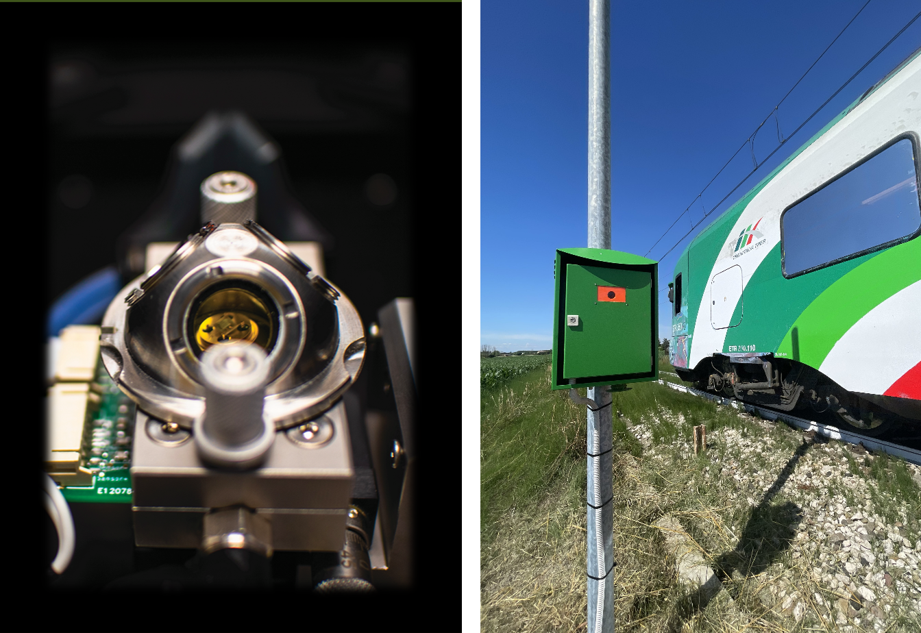 The final production phase of solid-state gas sensors involves packaging the substrates onto suitable supports that enable device characterization. The main activities carried out include:
- Characterization of the sensing properties of semiconductor films: study of the electrical and optical (UV-vis/NIR) characteristics of nanostructured semiconductor-based sensitive films produced in cleanrooms, exploring their behavior in response to various physical and chemical stimuli in dedicated test chambers, operating in photo- and thermo-activated modes. The sensing mechanisms are studied through analysis of solid-gas interface reactions using operando DRIFT spectroscopy.
- Application of solid-state gas sensors: fully fabricated devices at the DFST are implemented in olfactory systems designed for monitoring gaseous emissions in relevant indoor and outdoor environments, ranging from atmospheric pollutant control to air quality monitoring in academic classrooms and museum systems, from water resource management in precision agriculture to medical diagnostics of tumor pathologies.
Building C, Room C129
Contact persons: Prof. Vincenzo Guidi - Prof. Cesare Malagù - Dr. Barbara Fabbri - Dr. Giulia Zonta
Fabrication of Photovoltaic Devices
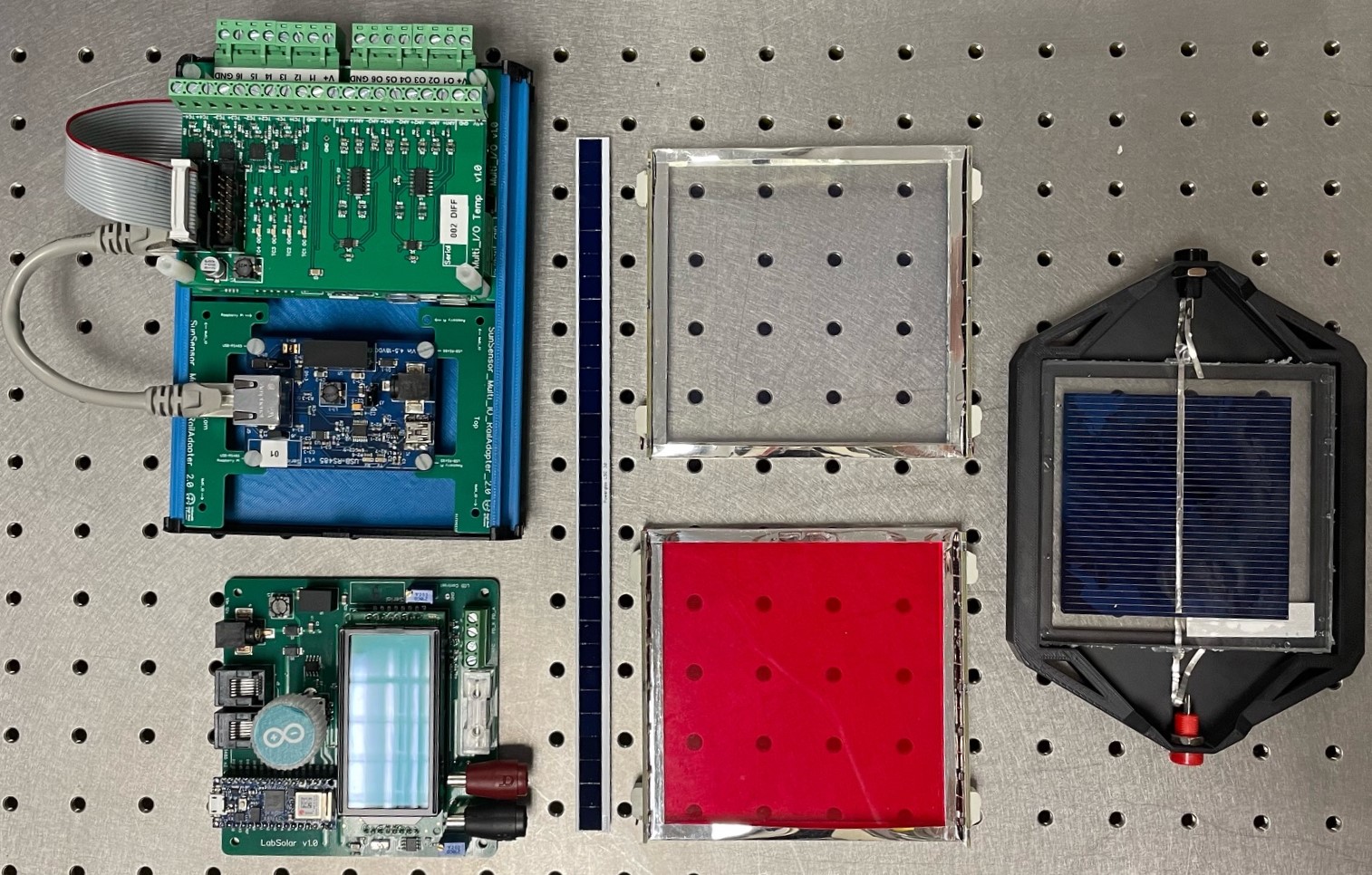 Design, testing, and development of photovoltaic devices and systems. Optical and thermal characterization and modeling of prototypes, both in controlled environments and operating conditions. Prototyping and assembly line for electronic boards using surface-mount technology (SMD).
Building C, Rooms C128 - C130 - C131
Contact person: Prof. Donato Vincenzi
Crystals
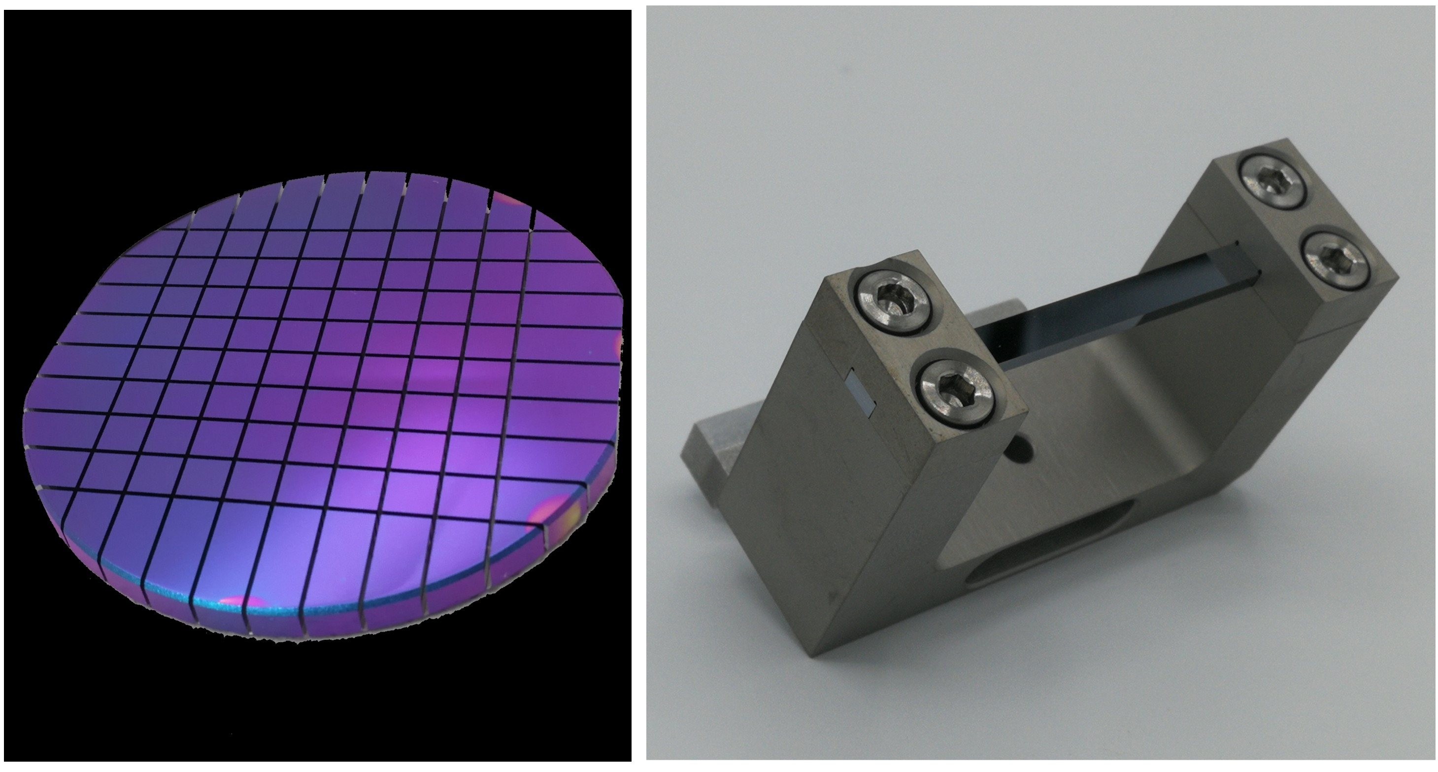 The Crystals Laboratory models semiconductor wafers into crystalline samples for various applications, including channeling in accelerators, development of innovative detectors for neutrinos and dark matter, and production of advanced optics for synchrotrons and XFEL. The laboratory is equipped with a computer numerical control (CNC) machine for automatic cutting and engraving semiconductors, glass, and ceramics, with micrometric precision. It also has a lapping and polishing station. Two 3D printers enable rapid production of objects in ABS and PLA.
Building C, Room C132
Contact Persons: Dr. Andrea Mazzolari - Dr. Marco Romagnoni
Spinlab
The Spinlab aboratory hosts a series of activities related to the spin physics of fundamental subnuclear constituents. It includes the following apparatus and technologies:
- Vacuum Technology: various pumping systems to meet the needs of the group primarily engaged in polarized nuclear targets, detectors and particle trackers with SiPMs and Cherenkov radiation; dry leak detectors for He and H₂ in both vacuum-testing and filling configurations (sniffers).
- Calibrated Flow Injection System: this system, with injection accuracies within one percent, enables (1) measurement of pumping speeds for gases relevant to polarized and non-polarized nuclear targets; (2) verification of conductances of accumulation cells, including openable cells used at HERMES (DESY-Hamburg), COSY (Jülich), and recently installed upstream of LHCb at CERN.
- Atomic Beams: the laboratory is equipped with an atomic beam and has hosted a polarized atomic beam, which was tested and adapted for use at PNPI in Gatchina (Russia). It plans to host the target used so far at COSY, adapting it for use at LHC in the LHC-Spin project.
- Accumulation Cell Characterization: equipped with a vacuum chamber for mechanical, thermal, and tightness characterization of accumulation cells for polarized nuclear targets. Cells designed and fabricated at Ferrara for various experiments have been characterized and tested using this chamber.
- Atomic Beam Diagnostics: the diagnostic system allows characterization of beam sources in terms of dissociation degree and intensity.
- Cryogenic Sensor and System Characterization Chamber: the lab hosts a vacuum chamber with an internal copper thermal shield and various cold heads. This chamber is used for (1) performance verification of dry cryogenic cooling systems with cold heads, (2) temperature sensor characterization and calibration, (3) Hall sensor characterization and calibration, and (4) testing and verification of contact cooling systems.
Contact person: Prof. Giuseppe Ciullo
Particle Detector Characterization System
The system enables measurements of Cherenkov radiation and the characterization and testing of scintillation detection and tracking systems for particles.
Thermal Treatments of Detectors
The laboratory is equipped with a series of ovens and furnaces designed for the thermal treatment of detectors.
Contact person: Dott. Marco Contalbrigo
Building G, Room G012
X-ray Diffractometry

X-ray diffraction occurs when electromagnetic waves with wavelengths in the order of angstroms (0.1 nm) are scattered with constructive interference by a crystalline material as defined by a periodic translation lattice. From the diffraction pattern recorded, it is possible to obtain information both on the unit cell (fundamental translations) that defines the crystal lattice and on the content (nature and position of the atoms) of the cell. The technique therefore allows a crystalline solid to be characterised at the atomic scale. When applied to polycrystalline samples, as in this specific case, it provides the following determinations in a very versatile way:
- identification of crystalline phases (‘qualitative analysis’) present in a polyphase sample even in minimal proportions (> 0.3-1 %) and with crystallite sizes well below nm;
- accurate determination of the weight percentages of crystalline phases and the amorphous phase contained in a polyphase mixture (Quantitative Phase Analysis, QPA)
- refinement of crystallographic parameters for the description of crystalline structures
- determination of microstructural parameters (crystallite sizes and lattice deformation)
- accurate identification of clay minerals on isotropic films.
For analyses b), c) and d), measurements are typically combined with diffraction pattern processing using the Rietveld method. This method consists of simulating the measured pattern using a pattern calculated on the basis of instrumental, structural and microstructural parameters optimised through least squares.
Some application topics: Removal of emerging pollutants using minerals and their synthetic analogues; Characterisation of gemmological materials; Design of binding materials and recycling of demolition waste; Energy optimisation of ceramic pigments for digital decoration; Property-structure relationship in sustainable catalysis; Stratigraphy studies and palaeoclimatic indications from clay minerals; Determination of expandable minerals in geotechnical engineering; Territorial connotation in viticulture through clay minerals; Applications in archaeometry (provenance, indirect dating, alteration products); Determination of harmful particulates in the workplace; Quantification of amorphous components in composite ceramics (e.g. glass-ceramics).
The X-ray Diffractometry Laboratory has the following devices:
- Bruker D8 Advance Da Vinci ‘powder’ diffractometer equipped with LynxEye-XT linear detector, which allows for the complete elimination of Kβ radiation. The following components are applicable: i) single sample holder for transmission measurements; ii) 9-position automatic sampler; iii) spinner for capillary tube that can be combined with a heating chamber up to 900°C; iv) automatic divergent slits or focusing mirror for grazing incidence measurements; v) collimator with ø 1 mm for point measurements (T07);
- Bruker D8 Advance powder diffractometer equipped with SOL-X point detector, with optimal energy resolution to eliminate Kβ and fluorescence radiation. Applicable: i) 9-position sampler; ii) chamber for controlled humidity measurements (T07);
- Mini ball mill and McCrone mill for controlled particle size reduction of powders;
- Millipore filtration system for preparing fine clay fraction samples in the form of films with crystallite orientation (T09).
Building B, Rooms T07 - T08 - T09 - T18
Contact persons: Prof. Giuseppe Cruciani - Prof.ssa Annalisa Martucci
